Area of density on mammogram
Home » Doctor Visit » Area of density on mammogramArea of density on mammogram
Area Of Density On Mammogram. Doctors who review mammograms are called radiologists. If a mammogram screening identifies developing asymmetry, there is a 12.8 percent chance that the person will develop breast cancer. If the shadow remains, then ultrasound may be done to determine if the area is solid or fluid filled (cystic). Interval change might be one area where a computer aided detection system can assist in.
 Scattered Fibroglandular Breast Tissue From medicalnewstoday.com
It is not associated with malignancy but the denseness makes it more difficult to read the mammogram and it is a way for the radiologist to say that the reading may be suboptimal but it can�t be helped. The breasts are extremely dense, which lowers the sensitivity of mammography. Scattered areas of fibroglandular density indicates there are some scattered areas of density, but the majority of the breast tissue is nondense. Breast density refers to the amounts of these different tissue types that are visible on a mammogram. Dear judy ro, it is not uncommon to need additional views after a screening mammogram. Scattered areas of fibroglandular density meaning some scattered areas are seen.
Interval change might be one area where a computer aided detection system can assist in.
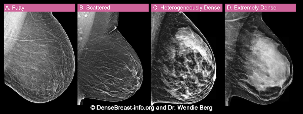
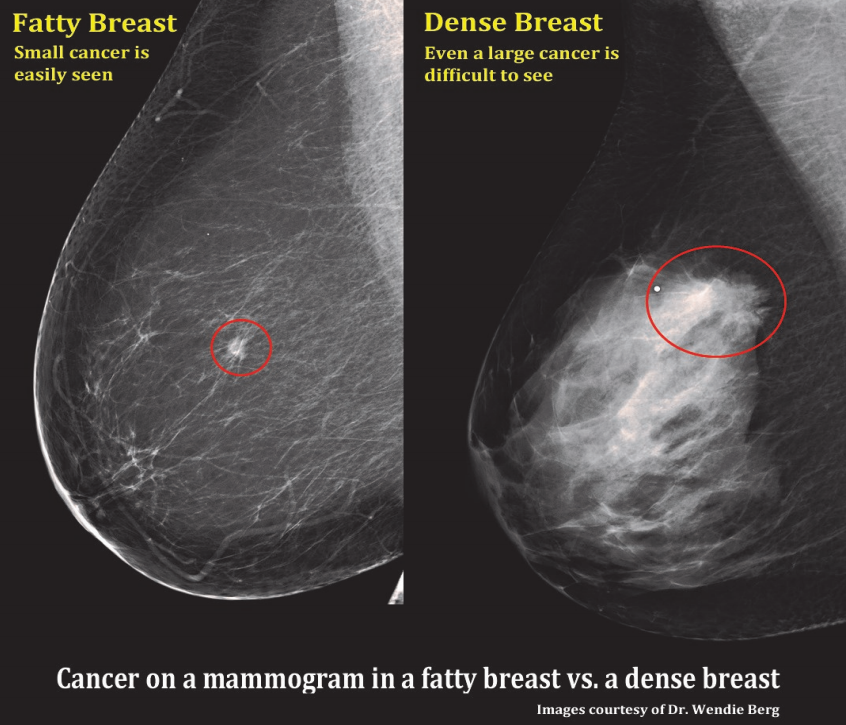
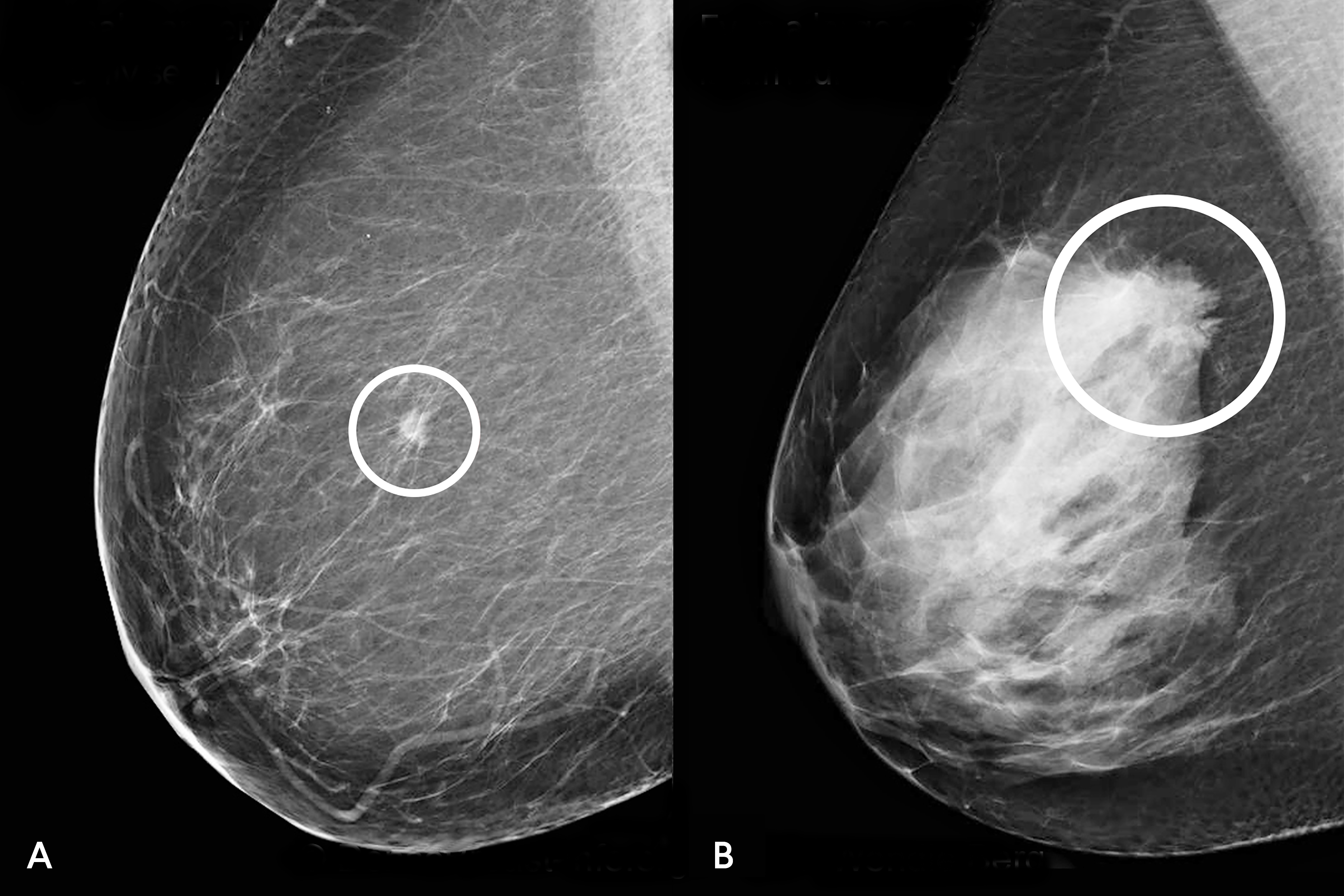
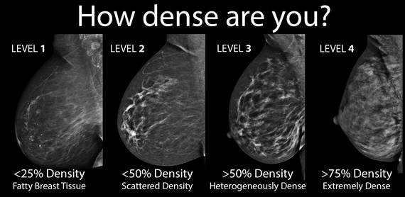
The radiologist decides which of the 4 categories best describes how dense your breasts are: If a cancer grows in an area of normal dense tissue, it can be hard or even impossible to see cancer on a mammogram. (a) almost entirely fatty breast tissue, found in about 10% of women. Breast density refers to the amounts of these different tissue types that are visible on a mammogram. Doctors who review mammograms are called radiologists. On a mammogram, an asymmetry typically means there’s more tissue, or white stuff on the mammogram, in one area than on the opposite side.
 Source: densebreast-info.org
Breast tissue can sometimes fold over on itself and cause a shadow (density) to appear. There has been a change since the prior mammogram, with an area of relatively denser tissue present in a new area. When asymmetry occurs, it leads to a question: A radiologist will examine a mammogram to look at the difference in position, volume and form of the breasts. The criteria for an asymmetry include that it is seen.
 Source: researchgate.net
The breasts are heterogeneously dense, which may obscure small masses. At this point this does not mean you have a mass, only a potential abnormality. About 4 in 10 women have this result. About 1 in 10 women has this result. On a mammogram, cancer is white.
 Source: moffitt.org
The criteria for an asymmetry include that it is seen. If a cancer grows in an area of normal dense tissue, it can be hard or even impossible to see cancer on a mammogram. More of the breast is made of dense glandular and fibrous tissue (described. A biopsy of these is essential. Up to 80% (but not 100%) of these masses are cancerous.
 Source: drattai.com
Have guidelines requiring that people be notified about their breast density after having a mammogram. About 1 in 10 women has this result. It is not definitely seen on the craniocaudal view. Rest of the fibroglandular tissue of both breasts shows no significant change. Breasts that are described as dense have more fibrous and glandular (fibroglandular) tissue.
 Source: kpwashingtonresearch.org
This is usually found in 10% of women. Almost entirely fatty meaning the breast are almost composed with fat. More of the breast is made of dense glandular and fibrous tissue (described. Otherwise, a certain amount of asymmetrical breast tissue is a normal variation that occurs in some women. There are scattered areas of fibroglandular density.
 Source: breast360.org
A radiologist will examine a mammogram to look at the difference in position, volume and form of the breasts. Have guidelines requiring that people be notified about their breast density after having a mammogram. This is usually found in 10% of women. If a mammogram screening identifies developing asymmetry, there is a 12.8 percent chance that the person will develop breast cancer. This needs additional evaluation, probably special mammographic views and maybe ultrasound.
 Source: researchgate.net
Up to 80% (but not 100%) of these masses are cancerous. Increased density usually refers to the presence of more glandular tissue than fat. An asymmetric density mammogram result is only a concern if there are associations with a clinically palpable abnormality, or a palpable mass. A biopsy of these is essential. (b) scattered areas of dense glandular tissue and fibrous connective tissue ( scattered fibroglandular breast tissue) found in about 40% of women.
 Source: itnonline.com
There has been a change since the prior mammogram, with an area of relatively denser tissue present in a new area. On a mammogram, cancer is white. An asymmetric density mammogram result is only a concern if there are associations with a clinically palpable abnormality, or a palpable mass. There are different kinds of asymmetries, from difference in size to tissue density. The breasts are almost entirely fatty.
 Source: breastcancer.org
Currently, 38 states and washington d.c. The breasts are almost entirely fatty. About 4 in 10 women have this result. Scattered areas of fibroglandular density indicates there are some scattered areas of density, but the majority of the breast tissue is nondense. Up to 80% (but not 100%) of these masses are cancerous.
 Source: breastlink.com
If a cancer grows in an area of normal dense tissue, it can be hard or even impossible to see cancer on a mammogram. Heterogeneously dense indicates that there are some. Scattered areas of fibroglandular density indicates there are some scattered areas of density, but the majority of the breast tissue is nondense. There are scattered areas of dense glandular and fibrous tissue (seen as white areas on the mammogram). The radiologist decides which of the 4 categories best describes how dense your breasts are:
 Source: mammalivefoundation.org
Breast density refers to the amounts of these different tissue types that are visible on a mammogram. It is not definitely seen on the craniocaudal view. Other possible causes for an asymmetrical breast density. If a cancer grows in an area of normal dense tissue, it can be hard or even impossible to see cancer on a mammogram. Breast density varies greatly by age and weight.
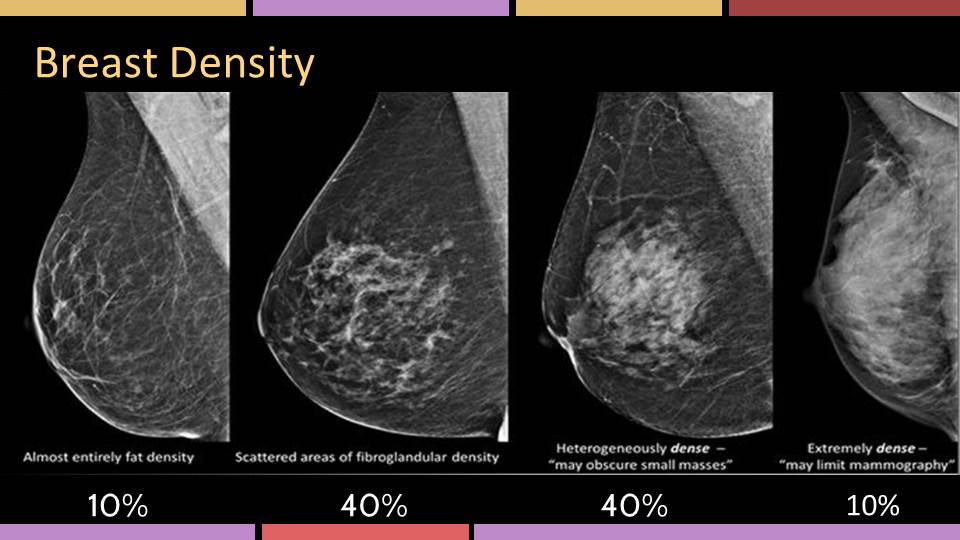
There are scattered areas of dense glandular and fibrous tissue (seen as white areas on the mammogram). Almost entirely fatty meaning the breast are almost composed with fat. Rest of the fibroglandular tissue of both breasts shows no significant change. An asymmetric density mammogram result is only a concern if there are associations with a clinically palpable abnormality, or a palpable mass. It is like trying to see a snowball in a blizzard.
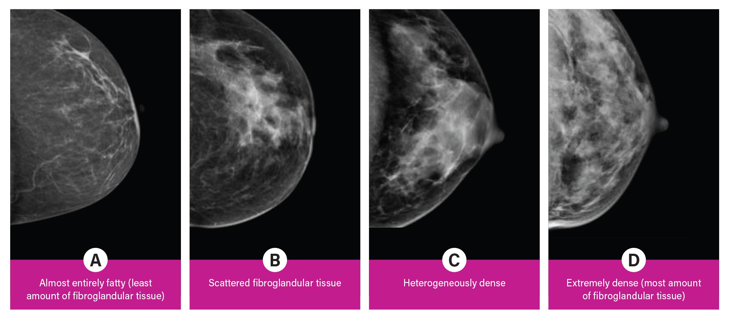 Source: itnonline.com
A new density in the left breast on the oblique view superiorly measuring 6 x 7 mm is 6.7 cm from the nipple. Doctors who review mammograms are called radiologists. The breasts are extremely dense, which lowers the sensitivity of mammography. Currently, 38 states and washington d.c. Rest of the fibroglandular tissue of both breasts shows no significant change.
 Source: researchgate.net
This is usually found in 10% of women. At this point this does not mean you have a mass, only a potential abnormality. In most cases, the breasts are generally symmetric in their density and architecture, but sometimes a report may reveal asymmetric density, which is common and usually noncancerous. About 4 in 10 women have this result. About 1 in 10 women has this result.
 Source: medicalnewstoday.com
(b) scattered areas of dense glandular tissue and fibrous connective tissue ( scattered fibroglandular breast tissue) found in about 40% of women. Rest of the fibroglandular tissue of both breasts shows no significant change. Breast density refers to the amounts of these different tissue types that are visible on a mammogram. It is not associated with malignancy but the denseness makes it more difficult to read the mammogram and it is a way for the radiologist to say that the reading may be suboptimal but it can�t be helped. Doctors who review mammograms are called radiologists.
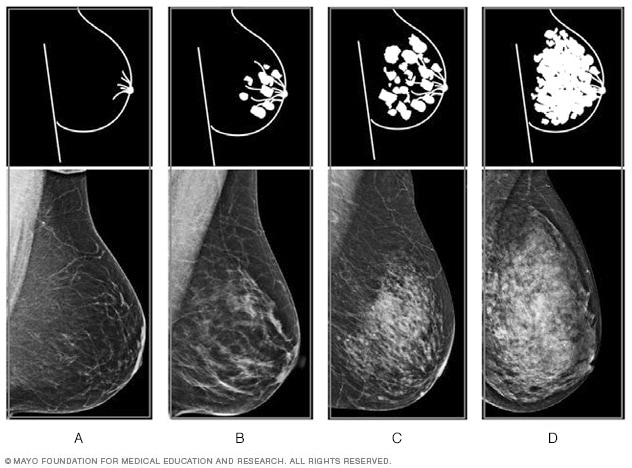 Source: mayoclinic.org
The criteria for an asymmetry include that it is seen. If a cancer (white) develops in an area of fat (black or dark gray/grey), it is usually easy to see. It is like trying to see a snowball in a blizzard. Breasts are almost all fatty tissue. On a mammogram, cancer is white.
 Source: keranews.org
On a mammogram, an asymmetry typically means there’s more tissue, or white stuff on the mammogram, in one area than on the opposite side. Scattered areas of fibroglandular density meaning some scattered areas are seen. Other possible causes for an asymmetrical breast density. In most cases, the breasts are generally symmetric in their density and architecture, but sometimes a report may reveal asymmetric density, which is common and usually noncancerous. This is usually found in 10% of women.
 Source: fredhutch.org
Interval change might be one area where a computer aided detection system can assist in. Heterogeneously dense indicates that there are some. On a mammogram, cancer is white. Both are features we look at on your breast imaging study. Almost entirely fatty meaning the breast are almost composed with fat.
If you find this site adventageous, please support us by sharing this posts to your favorite social media accounts like Facebook, Instagram and so on or you can also bookmark this blog page with the title area of density on mammogram by using Ctrl + D for devices a laptop with a Windows operating system or Command + D for laptops with an Apple operating system. If you use a smartphone, you can also use the drawer menu of the browser you are using. Whether it’s a Windows, Mac, iOS or Android operating system, you will still be able to bookmark this website.
Category
Related By Category
- Metastatic thyroid cancer prognosis
- Endocrinologist diabetes type 2
- How fast does colon cancer spread
- Hip replacement in elderly
- Physical therapy after arthroscopic shoulder surgery
- Symptoms of bacterial meningitis in children
- Chromophobe renal cell carcinoma
- Eye color change surgery usa
- Pradaxa vs eliquis vs xarelto
- Advanced stomach cancer symptoms