Biopsy for hair loss
Home » Doctor Visit » Biopsy for hair lossBiopsy for hair loss
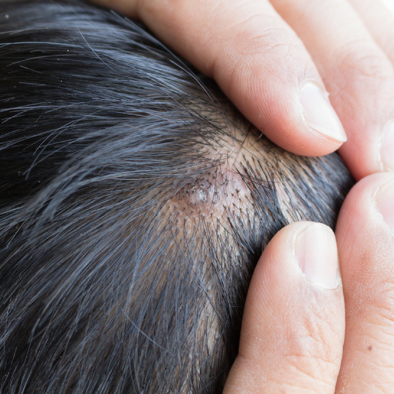
Biopsy For Hair Loss. In this procedure, a small portion of the scalp is removed from the head of the patient. The punch is 4 mm in diameter and it should be 4 mm deep so it includes follicular units and subcutaneous tissue. If hair loss does not resolve, a scalp biopsy to differentiate between alopecia areata, telogen effluvium, and male or female pattern hair loss should. The precise follow up time depends on the type of biopsy you are having and specifics to the type of hair loss condition.
 I Got A Scalp Biopsy | Hair Loss Journey - Youtube From youtube.com
Repetitive stress on the hair can cause chronic alopecia and damage the hair shaft and hair follicles. Generally speaking, a scalp biopsy is likely recommended if the patient suffers hair loss without any particular cause. The procedure takes about five minutes and is primarily used to help determine the cause of hair loss. You may dye and colour your hair one week after the biopsy. We can use the results of a biopsy to make or confirm a diagnosis of alopecia. Product can be used on the scalp the very next day.
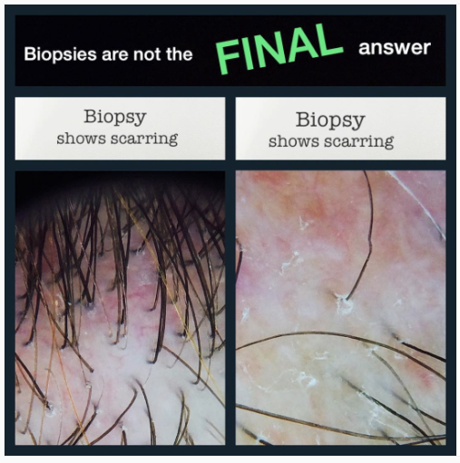
When a follicle scarring process appears to be the cause of a patient’s hair loss, a scalp biopsy is often necessary to establish or confirm a diagnosis.
We can also obtain important information in cases of unexplained hair loss or when the potential for regrowth. Generally speaking, a scalp biopsy is likely recommended if the patient suffers hair loss without any particular cause. In this procedure, a small portion of the scalp is removed from the head of the patient. Your doctor scrapes samples from the skin or from a few hairs plucked from the scalp to examine the hair roots under a microscope. It is important to identify the normal hair follicle structure, the number, size and distribution of hair follicles within a biopsy specimen. A punch biopsy is preferred and is easily done at the.
 Source: youtube.com
There are many hair loss conditions that resemble each other and a biopsy is helpful to differentiate between these mimickers. Repetitive stress on the hair can cause chronic alopecia and damage the hair shaft and hair follicles. This physical damage to the hair shaft like tight braids and ponytails, excessive brushing, using rollers and straightening. You may dye and colour your hair one week after the biopsy. For example, some forms of lichen planopilaris (a scarring hair loss condition) can look nearly identical to some forms of androgenetic alopecia (a non scarring hair loss condition).
 Source: sciencedirect.com
How a scalp biopsy for hair loss performed. For example, some forms of lichen planopilaris (a scarring hair loss condition) can look nearly identical to some forms of androgenetic alopecia (a non scarring hair loss condition). The tissue is then sent to a dermatopathologist for further analysis. A follow up appointment via phone will be arranged 4 weeks after your appointment. Repetitive stress on the hair can cause chronic alopecia and damage the hair shaft and hair follicles.
 Source: intechopen.com
You may dye and colour your hair one week after the biopsy. For example, some forms of lichen planopilaris (a scarring hair loss condition) can look nearly identical to some forms of androgenetic alopecia (a non scarring hair loss condition). The tissue is then sent to a dermatopathologist for further analysis. The histological findings in many forms of hair loss may be similar, and an accurate diagnosis of hair loss depends on distinguishing abnormal from normal follicular architecture. Therefore,careful selection of the scalp biopsy site, optimal biopsy technique, and proper specimen processing followed by informed histological interpretation are essential steps in the successful evaluation of both cicatricial and.

The biopsy does require local anesthetic, with most individuals. It is performed under local anesthesia. Prp for hair loss other hair treatments village dermatology houston. It is important to identify the normal hair follicle structure, the number, size and distribution of hair follicles within a biopsy specimen. This can help determine whether an infection is causing hair loss.
 Source: donovanmedical.com
If hair loss does not resolve, a scalp biopsy to differentiate between alopecia areata, telogen effluvium, and male or female pattern hair loss should. Hair in the frontal region become thin and sparse. How a scalp biopsy for hair loss performed. Your doctor scrapes samples from the skin or from a few hairs plucked from the scalp to examine the hair roots under a microscope. Therefore,careful selection of the scalp biopsy site, optimal biopsy technique, and proper specimen processing followed by informed histological interpretation are essential steps in the successful evaluation of both cicatricial and.

In this procedure, a small portion of the scalp is removed from the head of the patient. Your doctor uses a special instrument to examine hairs trimmed at their bases. I believe that i am in the early process of hair loss. That portion is then examined by a dermatologist under a microscope to determine what’s causing hair loss. Product can be used on the scalp the very next day.
 Source: researchgate.net
Prp for hair loss other hair treatments village dermatology houston. The histological findings in many forms of hair loss may be similar, and an accurate diagnosis of hair loss depends on distinguishing abnormal from normal follicular architecture. I believe that i am in the early process of hair loss. To determine the cause of hair loss, your dermatologist asks a variety of questions about when hair loss. When your doctor performs a scalp biopsy, he or she will take a small section of your scalp, usually about 4mm in diameter, which is removed and examined under a microscope.
 Source: flickr.com
The histological findings in many forms of hair loss may be similar, and an accurate diagnosis of hair loss depends on distinguishing abnormal from normal follicular architecture. In this procedure, a small portion of the scalp is removed from the head of the patient. Product can be used on the scalp the very next day. In this procedure, a small portion of the scalp is removed from the head of the patient. Repetitive stress on the hair can cause chronic alopecia and damage the hair shaft and hair follicles.
 Source: ishrs-htforum.org
The dermatologist will first clean and disinfect the area that needs to be biopsied and then mark it. If hair loss does not resolve, a scalp biopsy to differentiate between alopecia areata, telogen effluvium, and male or female pattern hair loss should. In this procedure, a small portion of the scalp is removed from the head of the patient. When your doctor performs a scalp biopsy, he or she will take a small section of your scalp, usually about 4mm in diameter, which is removed and examined under a microscope. This physical damage to the hair shaft like tight braids and ponytails, excessive brushing, using rollers and straightening.
 Source: jaad.org
The dermatologist will first clean and disinfect the area that needs to be biopsied and then mark it. How a scalp biopsy for hair loss performed. A follow up appointment via phone will be arranged 4 weeks after your appointment. The precise follow up time depends on the type of biopsy you are having and specifics to the type of hair loss condition. We can also obtain important information in cases of unexplained hair loss or when the potential for regrowth.
 Source: youtube.com
The tissue is then sent to a dermatopathologist for further analysis. The dermatologist will first clean and disinfect the area that needs to be biopsied and then mark it. The punch is 4 mm in diameter and it should be 4 mm deep so it includes follicular units and subcutaneous tissue. How a scalp biopsy for hair loss performed. For example, some forms of lichen planopilaris (a scarring hair loss condition) can look nearly identical to some forms of androgenetic alopecia (a non scarring hair loss condition).
 Source: donovanmedical.com
My biopsy came back with androgenetic alopecia and te. Learn more about hair loss treatments: When your doctor performs a scalp biopsy, he or she will take a small section of your scalp, usually about 4mm in diameter, which is removed and examined under a microscope. In this procedure, a small portion of the scalp is removed from the head of the patient. No one in my family has female pattern baldness.
 Source: hairguard.com
I believe that i am in the early process of hair loss. The procedure takes about five minutes and is primarily used to help determine the cause of hair loss. When a follicle scarring process appears to be the cause of a patient’s hair loss, a scalp biopsy is often necessary to establish or confirm a diagnosis. Therefore,careful selection of the scalp biopsy site, optimal biopsy technique, and proper specimen processing followed by informed histological interpretation are essential steps in the successful evaluation of both cicatricial and. This can help determine whether an infection is causing hair loss.
 Source: thelondonskinandhairclinic.com
In this procedure, a small portion of the scalp is removed from the head of the patient. If hair loss does not resolve, a scalp biopsy to differentiate between alopecia areata, telogen effluvium, and male or female pattern hair loss should. When a follicle scarring process appears to be the cause of a patient’s hair loss, a scalp biopsy is often necessary to establish or confirm a diagnosis. A punch biopsy is preferred and is easily done at the. Your doctor uses a special instrument to examine hairs trimmed at their bases.
 Source: donovanmedical.tumblr.com
Hair loss,some times in other hair in my all of body and thin hair in all body some times loss eyelash and eyebrow reduce hair grow extreme muscle aches and pain. The dermatologist will first clean and disinfect the area that needs to be biopsied and then mark it. Hair loss,some times in other hair in my all of body and thin hair in all body some times loss eyelash and eyebrow reduce hair grow extreme muscle aches and pain. The precise follow up time depends on the type of biopsy you are having and specifics to the type of hair loss condition. This can help determine whether an infection is causing hair loss.
 Source: intechopen.com
We can use the results of a biopsy to make or confirm a diagnosis of alopecia. Your doctor uses a special instrument to examine hairs trimmed at their bases. To determine the cause of hair loss, your dermatologist asks a variety of questions about when hair loss. The precise follow up time depends on the type of biopsy you are having and specifics to the type of hair loss condition. I believe that i am in the early process of hair loss.
 Source: miamihair.com
The punch is 4 mm in diameter and it should be 4 mm deep so it includes follicular units and subcutaneous tissue. Product can be used on the scalp the very next day. We can use the results of a biopsy to make or confirm a diagnosis of alopecia. My biopsy came back with androgenetic alopecia and te. All of these diagnostic tests can be conducted in your dermatologist’s office.
 Source: propath.com
When a follicle scarring process appears to be the cause of a patient’s hair loss, a scalp biopsy is often necessary to establish or confirm a diagnosis. We can also obtain important information in cases of unexplained hair loss or when the potential for regrowth. It is performed under local anesthesia. You may dye and colour your hair one week after the biopsy. Hair loss,some times in other hair in my all of body and thin hair in all body some times loss eyelash and eyebrow reduce hair grow extreme muscle aches and pain.
If you find this site good, please support us by sharing this posts to your favorite social media accounts like Facebook, Instagram and so on or you can also save this blog page with the title biopsy for hair loss by using Ctrl + D for devices a laptop with a Windows operating system or Command + D for laptops with an Apple operating system. If you use a smartphone, you can also use the drawer menu of the browser you are using. Whether it’s a Windows, Mac, iOS or Android operating system, you will still be able to bookmark this website.
Category
Related By Category
- Metastatic thyroid cancer prognosis
- Endocrinologist diabetes type 2
- How fast does colon cancer spread
- Hip replacement in elderly
- Physical therapy after arthroscopic shoulder surgery
- Symptoms of bacterial meningitis in children
- Chromophobe renal cell carcinoma
- Eye color change surgery usa
- Pradaxa vs eliquis vs xarelto
- Advanced stomach cancer symptoms