Heart valves auscultation points
Home » Doctor Visit » Heart valves auscultation pointsHeart valves auscultation points
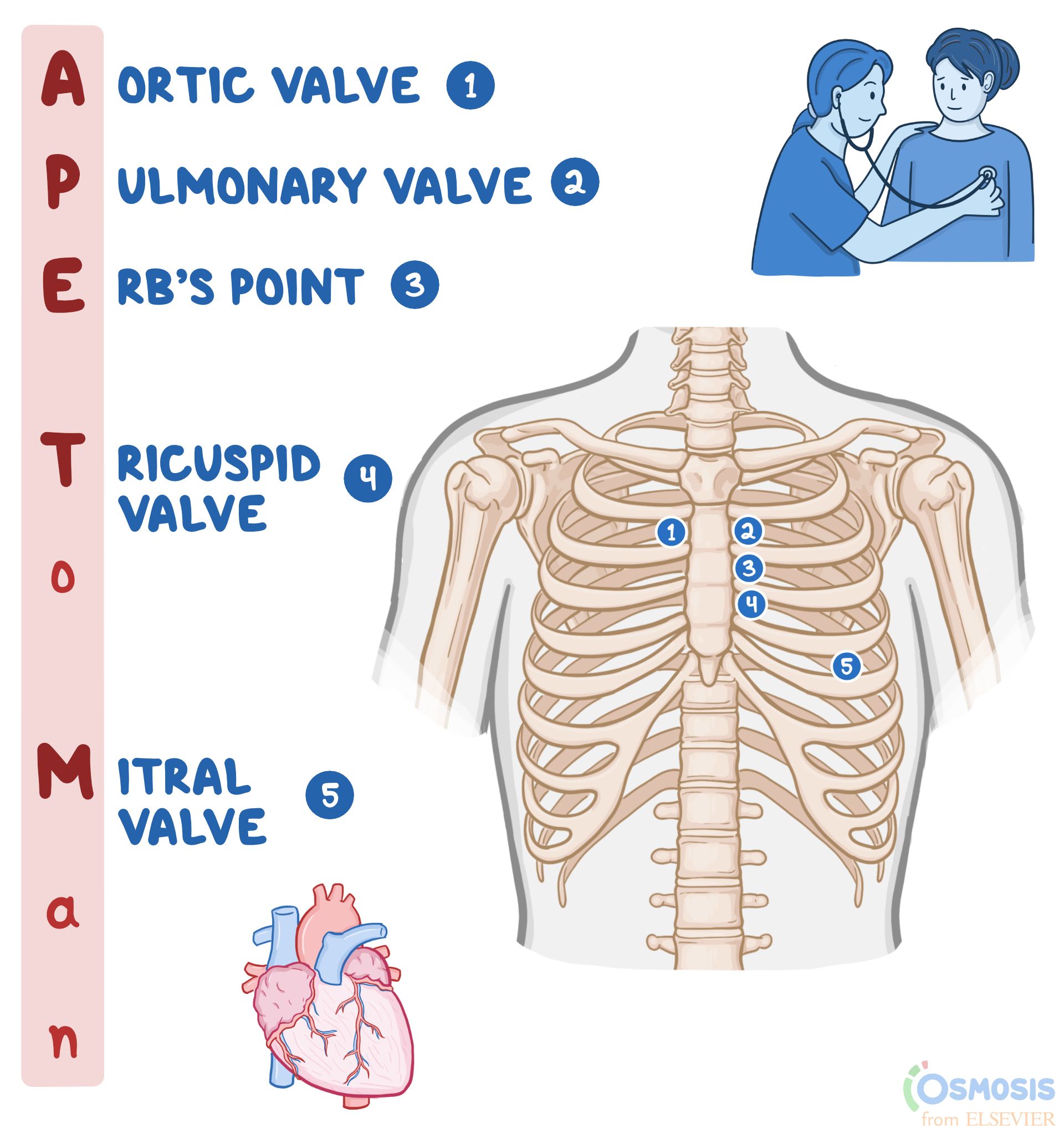
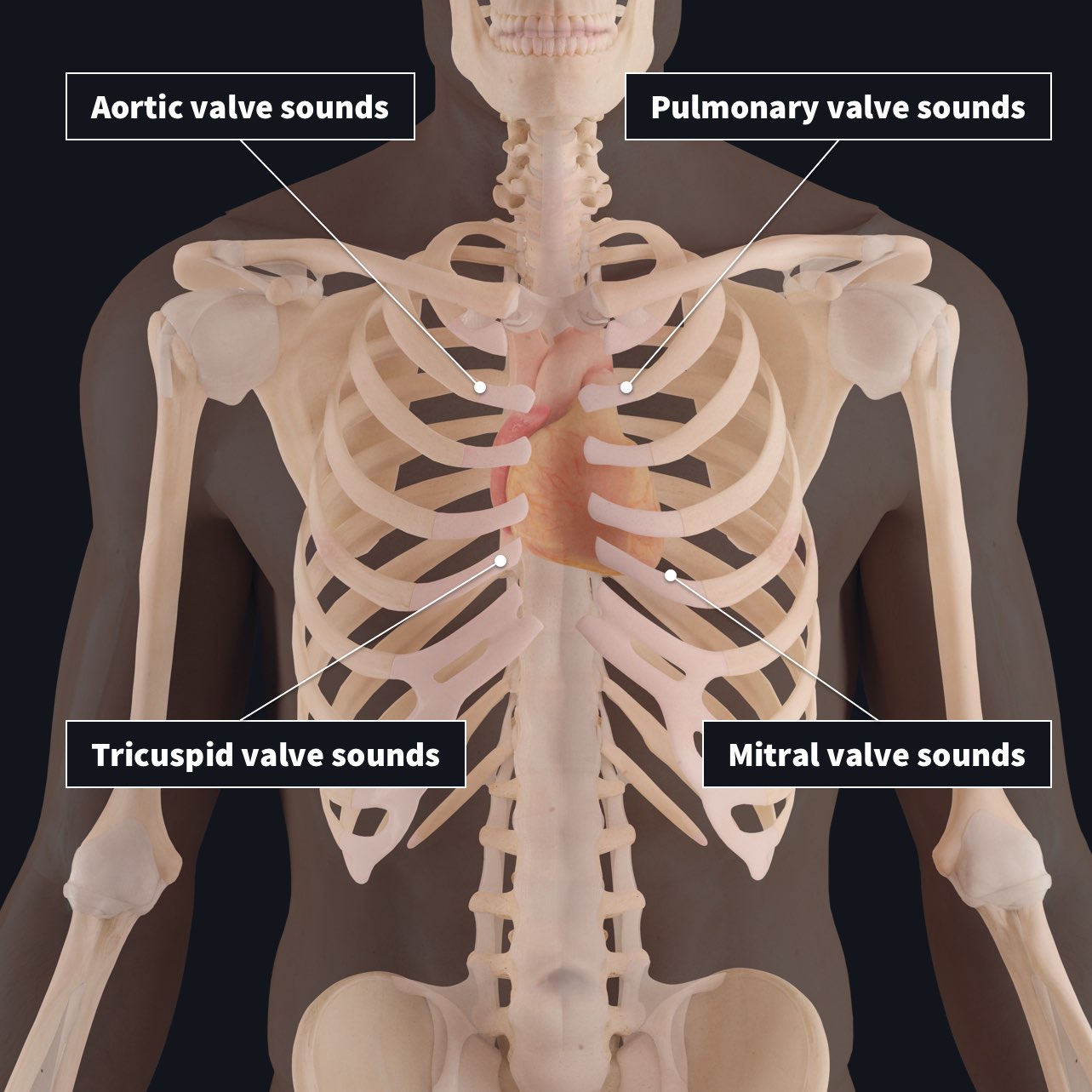
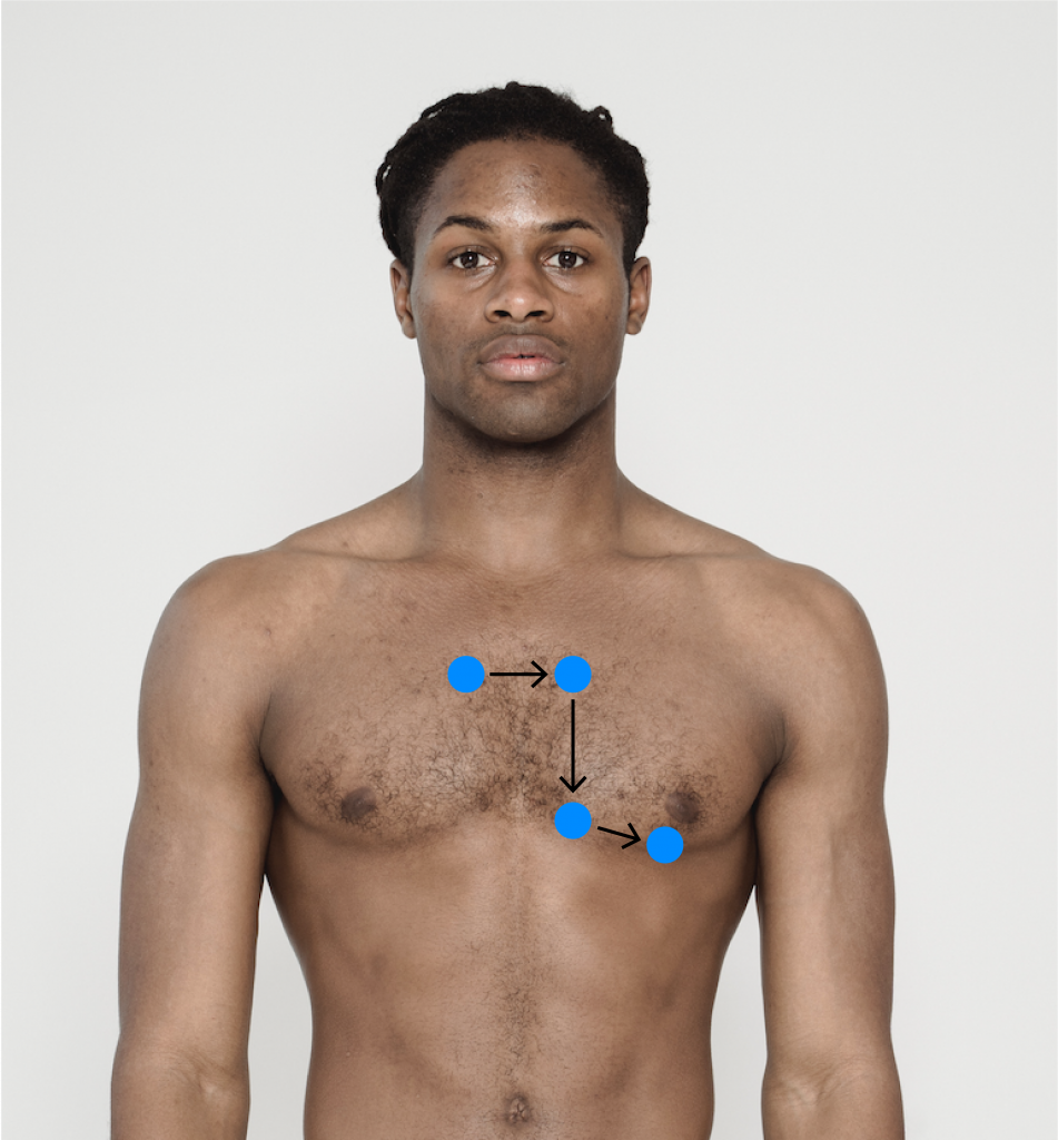
Heart Valves Auscultation Points. Your healthcare provider will listen for: The four standard points of auscultation for the heart are: 1 to compound this problem further. The nurse will be assessing s1 and s2 while noting if there are any s1 and s2 splits or extra heart sounds like s3, s4, or heart murmurs.
 Heart Sounds - Wikipedia From en.wikipedia.org
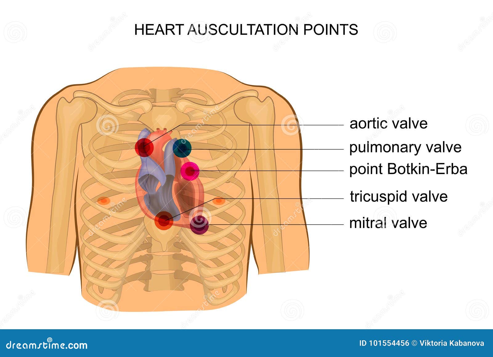
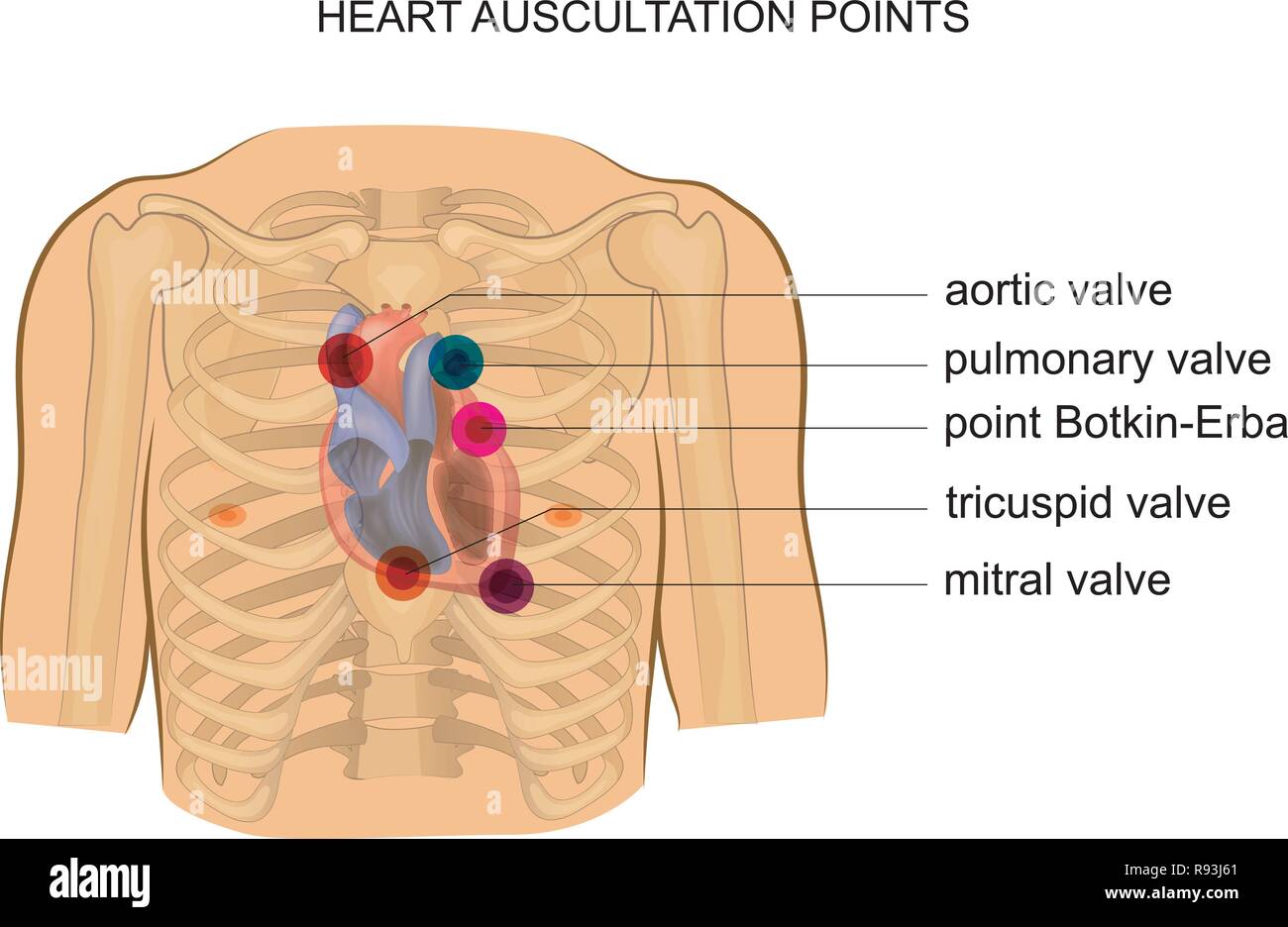
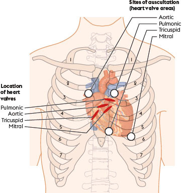
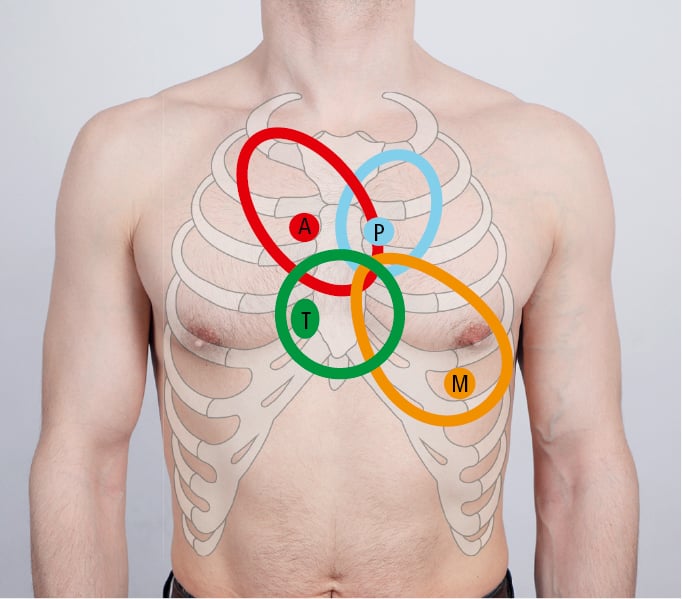
With the advent of numerous new diagnostic modalities, however, especially ultrasonic imaging and doppler techniques, cardiac auscultation is receiving less emphasis in teaching and practice. 1 to compound this problem further. All valves are a bit lower when the patient is standing. In older individuals it indicates the presence of congestive heart failure or valve disease. The pulmonary and aortic valves are both best heard in the 2nd intercostal space, to the left and right respectively. The 5 points of auscultation of the heart include the aortic, pulmonic, tricuspid, and mitral valve as well as an area called erb’s point, where s2 is best heard.
Vibrations of these structures from the blood flow create audible sounds — the more turbulent the blood flow, the more vibrations that get created.
Auscultation of the heart technique •to obtain the most information from cardiac auscultation and to assess correctly the findings, it is necessary to know the sites of valves projection on the chest wall and listening points of the heart. The locations of auscultation center around the heart valves. Keep the client in a supine position and use draping. Auscultation is performed for the purposes of examining the circulatory system and respiratory system (heart sounds and breath sounds), as well as the gastrointestinal system (bowel sounds). The 5 points of auscultation of the heart include the aortic, pulmonic, tricuspid, and mitral valve as well as an area called erb’s point, where s2 is best heard. A third heart sound occurs early in diastole.
 Source: gastroepato.it
As a result, the auscultatory areas of the heart valves are the following: Auscultation is the term for listening to the internal sounds of the body, usually using a stethoscope. Auscultating the heart allows the nurse to assess the heart’s rhythm, rate, and sound of valve closure. In older individuals it indicates the presence of congestive heart failure or valve disease. The aortic, pulmonic, tricuspid, and mitral valves are four of the five points of auscultation.
 Source: osmosis.org
Aortic valve th= left 4 intercostal space dorsal to the mv (usually the level of the point of the shoulder). It corresponds to the beginning of ventricular systole (waugh and grant, 2006). In young people and athletes it is a normal phenomenon. First heart sounds heard better at mv. As a result, the auscultatory areas of the heart valves are the following:
 Source: researchgate.net
This can make differentiation quite difficult. 1 to compound this problem further. Auscultation of the heart technique •to obtain the most information from cardiac auscultation and to assess correctly the findings, it is necessary to know the sites of valves projection on the chest wall and listening points of the heart. Auscultation is a method used to listen to the sounds of your body during a physical examination by using a stethoscope. What point of auscultation is the true projection of the heart valves?
 Source: amboss.com
For most of this century, the stethoscope has served as a critical diagnostic tool in cardiovascular evaluation. Your healthcare provider will listen for: The fifth is erb’s point, located left of the sternal border in the third intercostal space. 3) with the heart superimposed. A patient’s lungs, heart, and intestines are the most common organs heard during auscultation.
 Source: 3d4medical.com
Aortic valve th= left 4 intercostal space dorsal to the mv (usually the level of the point of the shoulder). It is very important you are able to understand how to distinguish between s1 and s2 and what s3, s4. The stethoscope is an instrument that does not significantly amplify sound, but, more important, acts as a selective filter of. In young people and athletes it is a normal phenomenon. Similar to each component of an exam, auscultation is only a piece of the puzzle and should be used in combination with other findings.
 Source: pressbooks.library.torontomu.ca
In young people and athletes it is a normal phenomenon. A third heart sound occurs early in diastole. The locations of auscultation center around the heart valves. Second heart sound heard better at aov and pv. In young people and athletes it is a normal phenomenon.
 Source: dreamstime.com
For most of this century, the stethoscope has served as a critical diagnostic tool in cardiovascular evaluation. It corresponds to the beginning of ventricular systole (waugh and grant, 2006). For most of this century, the stethoscope has served as a critical diagnostic tool in cardiovascular evaluation. With the advent of numerous new diagnostic modalities, however, especially ultrasonic imaging and doppler techniques, cardiac auscultation is receiving less emphasis in teaching and practice. The second sound is called s2, or a dub.
 Source: youtube.com
In young people and athletes it is a normal phenomenon. The four standard points of auscultation for the heart are: It is an integral part of physical examination of a. Keep the client in a supine position and use draping. The fifth is erb’s point, located left of the sternal border in the third intercostal space.
 Source: pinterest.com
The nurse will be assessing s1 and s2 while noting if there are any s1 and s2 splits or extra heart sounds like s3, s4, or heart murmurs. The same variables determine the turbulence of blood flow as all fluids. A patient’s lungs, heart, and intestines are the most common organs heard during auscultation. Your healthcare provider will listen for: The third heart sound.is caused by a sudden deceleration of blood flow into the left ventricle from the left atrium.
 Source: quizlet.com
It corresponds to the beginning of ventricular systole (waugh and grant, 2006). The fifth is erb’s point, located left of the sternal border in the third intercostal space. These heart auscultation points are all in your upper left chest area. The point of the elbow. Auscultation is the term for listening to the internal sounds of the body, usually using a stethoscope.
 Source: researchgate.net
All valves are a bit lower when the patient is standing. The 5 points of auscultation of the heart include the aortic, pulmonic, tricuspid, and mitral valve as well as an area called erb’s point, where s2 is best heard. When auscultating the heart which valves are heard best? Aortic valve th= left 4 intercostal space dorsal to the mv (usually the level of the point of the shoulder). 3) with the heart superimposed.
 Source: alamy.com
Sound from the aortic valve is often transmitted. Pulmonic valve = left 3rd intercostal. It is an integral part of physical examination of a. The first heart sound (‘lub’), which is often referred to as s1, is due to closure of the mitral and tricuspid valves and is best heard at the apex. When auscultating the heart which valves are heard best?
 Source: en.wikipedia.org
Second heart sound heard better at aov and pv. Cardiac auscultation is one of the keys to an effective physical exam and can often assist in the assessment of a patient’s hemodynamic function. Your healthcare provider can hear these sounds when your heart valves are closing. For most of this century, the stethoscope has served as a critical diagnostic tool in cardiovascular evaluation. A third heart sound occurs early in diastole.
 Source: journals.rcni.com
The pulmonary and aortic valves are both best heard in the 2nd intercostal space, to the left and right respectively. The second sound is called s2, or a dub. In young people and athletes it is a normal phenomenon. When auscultating the heart which valves are heard best? In older individuals it indicates the presence of congestive heart failure or valve disease.
 Source: empendium.com
The locations of auscultation center around the heart valves. Keep the client in a supine position and use draping. You can do this assessment with the client in a sitting position, but it is generally easier to do in a supine position because you can drape the client without struggling to hold the drape in place. The four standard points of auscultation for the heart are: 3) with the heart superimposed.
 Source: quizlet.com
The 5 points of auscultation of the heart include the aortic, pulmonic, tricuspid, and mitral valve as well as an area called erb’s point, where s2 is best heard. Heart sounds are created from blood flowing through the heart chambers as the cardiac valves open and close during the cardiac cycle. Keep the client in a supine position and use draping. The third heart sound.is caused by a sudden deceleration of blood flow into the left ventricle from the left atrium. As a result, the auscultatory areas of the heart valves are the following:

In older individuals it indicates the presence of congestive heart failure or valve disease. Vibrations of these structures from the blood flow create audible sounds — the more turbulent the blood flow, the more vibrations that get created. The four standard points of auscultation for the heart are: I sound components atrial valvular muscular. The point of the elbow.
 Source: easyauscultation.com
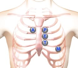
The four standard points of auscultation for the heart are: All valves are a bit lower when the patient is standing. As a result, the auscultatory areas of the heart valves are the following: The four standard points of auscultation for the heart are: Heart sounds are created from blood flowing through the heart chambers as the cardiac valves open and close during the cardiac cycle.
If you find this site good, please support us by sharing this posts to your own social media accounts like Facebook, Instagram and so on or you can also bookmark this blog page with the title heart valves auscultation points by using Ctrl + D for devices a laptop with a Windows operating system or Command + D for laptops with an Apple operating system. If you use a smartphone, you can also use the drawer menu of the browser you are using. Whether it’s a Windows, Mac, iOS or Android operating system, you will still be able to bookmark this website.
Category
Related By Category
- Metastatic thyroid cancer prognosis
- Endocrinologist diabetes type 2
- How fast does colon cancer spread
- Hip replacement in elderly
- Physical therapy after arthroscopic shoulder surgery
- Symptoms of bacterial meningitis in children
- Chromophobe renal cell carcinoma
- Eye color change surgery usa
- Pradaxa vs eliquis vs xarelto
- Advanced stomach cancer symptoms