Mri of brain and orbits
Home » Doctor Visit » Mri of brain and orbitsMri of brain and orbits
Mri Of Brain And Orbits. Under the control of mri, additional diagnostic tests can be performed, for example, a biopsy if there is a suspicion of a malignant tumor process inside the eye. It enables clinicians to focus on various parts of the brain and examine their anatomy and pathology, using different mri sequences, such as t1w, t2w, or flair. This article is intended to outline some general principles of protocol design. Angle the position block parallel to the rt and lt eye lenses.
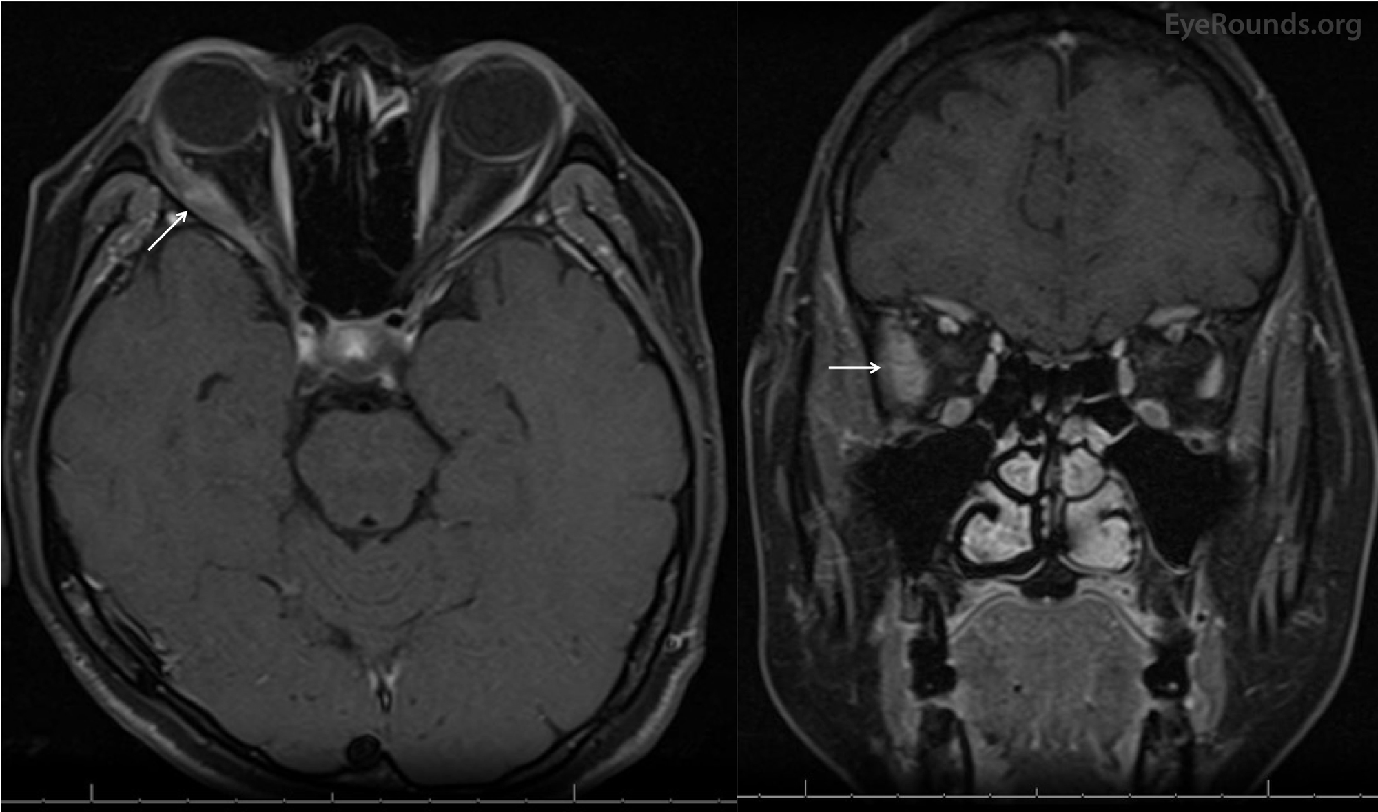
 Localized Orbital Amyloidosis Presenting as NewOnset Diplopia From webeye.ophth.uiowa.edu
The dural venous sinuses enhance normally. Mri brain and eye (orbits) magnetic resonance imaging (mri) is a safe as well as painless procedure. Other reasons a doctor might request an mri of the orbits are swelling, infection, or cellulitis near the eyes. An appropriate angle must be given in the sagittal plane on a tilted head (perpendicular to optic nerve). Mri brain is a specialist investigation that is used for the assessment of a number of neurological conditions. No abnormal gradient or diffusion signal is identified.
Check the positioning block in the other two planes.
Check the positioning block in the other two planes. This is another plus of the modern diagnostic method. Mri brain orbits what to expect when you arrive: At the same time, the potential risk associated with residual gadolinium concentrations in the brain should be taken into consideration.”(2) aim: To evaluate the usefulness of brain mri as compared to orbital mri in the assessment of idiopathic intracranial hypertension (iih). The dural venous sinuses enhance normally.
 Source: researchgate.net
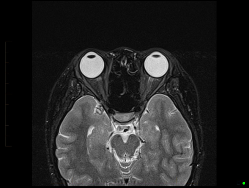
The orbit mri is similar to the brain mri with additional images specific to the eyes. An appropriate angle must be given in the sagittal plane on a tilted head (perpendicular to optic nerve). The specifics will vary depending on mri hardware and software, radiologist�s and referrer�s preference, institutional protocols, patient. With contrast, there is no abnormal enhancing lesion. An mri of the head and orbits was performed for 42 patients with the clinical diagnosis.
 Source: researchgate.net
If you have been booked for an orbit mri you will be asked to arrive at the mri 15 minutes prior to your appointment time. The orbits, in this context, are the eyes and structures surrounding them. Would a contrast mri of brain and orbits on a 1.5t machine be adequate to rule out a tumor in the brain stem? With contrast, there is no abnormal enhancing lesion. He was sent to mri because both of his optic nerves were swollen.
 Source: researchgate.net
A brain mri is one of the most commonly performed techniques of medical imaging. Mris of the orbit are also done when a patient has a bulging eye or eyes so the doctor can see if there is a mass or tumor in the area. Other reasons a doctor might request an mri of the orbits are swelling, infection, or cellulitis near the eyes. A brain mri is one of the most commonly performed techniques of medical imaging. A magnetic resonance imaging (mri) orbit scan will show:
 Source: researchgate.net
This is another plus of the modern diagnostic method. The aims of this retrospective study are: These studies help to detect abnormalities such as cysts, tumors, ms (multiple sclerosis), seizure, stroke and other. The specifics will vary depending on mri hardware and software, radiologist�s and referrer�s preference, institutional protocols, patient. Previously he was diagnosed with birdshot chorioretinopathy.
Source: researchgate.net
No abnormal gradient or diffusion signal is identified. Depending on what your doctor is looking for, this test may be ordered with or without iv contrast. Mri uses a magnetic field and radio waves to create detailed images of the organs and tissues within your body without the use of ionizing radiation. Mri brain and eye (orbits) magnetic resonance imaging (mri) is a safe as well as painless procedure. No abnormal enhancement is seen.
 Source: researchgate.net
Orbits protocol is an mri protocol comprising a group of mri sequences as a useful approach to routinely assess the orbits and their related conditions. Anterior margin of globe to anterior surface of pons, perpendicular to axis of the orbital segment of the optic nerves. Under the control of mri, additional diagnostic tests can be performed, for example, a biopsy if there is a suspicion of a malignant tumor process inside the eye. The optic nerves are symmetric and unremarkable. There is no enhancement following contrast administration.
 Source: researchgate.net
Mri brain and eye (orbits) magnetic resonance imaging (mri) is a safe as well as painless procedure. An appropriate angle must be given in the sagittal plane on a tilted head (perpendicular to optic nerve). Anterior margin of globe to anterior surface of pons, perpendicular to axis of the orbital segment of the optic nerves. Mri is used to analyze the anatomy of the brain and to identify some pathological. If you have been booked for an orbit mri you will be asked to arrive at the mri 15 minutes prior to your appointment time.
 Source: researchgate.net
Mri brain orbits what to expect when you arrive: Would a contrast mri of brain and orbits on a 1.5t machine be adequate to rule out a tumor in the brain stem? Mri uses a magnetic field and radio waves to create detailed images of the organs and tissues within your body without the use of ionizing radiation. When you arrive in the mri. It enables clinicians to focus on various parts of the brain and examine their anatomy and pathology, using different mri sequences, such as t1w, t2w, or flair.
 Source: researchgate.net
No abnormal enhancement is seen. This article is intended to outline some general principles of protocol design. Mri brain and eye (orbits) magnetic resonance imaging (mri) is a safe as well as painless procedure. Plan the coronal slices on the axial plane; Mr brain and orbits wo neuro protocol.
 Source: researchgate.net
What is a mri of the head (brain, iacs, orbits, pituitary) and what does it do? My husband had brain + orbits mri with / whithout contrast. Angle the position block parallel to the rt and lt eye lenses. An mri of the head and orbits was performed for 42 patients with the clinical diagnosis. What is a mri of the head (brain, iacs, orbits, pituitary) and what does it do?
 Source: researchgate.net
There is no enhancement following contrast administration. My husband had brain + orbits mri with / whithout contrast. He was sent to mri because both of his optic nerves were swollen. Orbits protocol is an mri protocol comprising a group of mri sequences as a useful approach to routinely assess the orbits and their related conditions. Mr brain and orbits wo neuro protocol.
 Source: dreamstime.com
The specifics will vary depending on mri hardware and software, radiologist�s and referrer�s preference, institutional protocols, patient. The orbits, paranasal sinuses and mastoid air cells are. This mri orbits and paranasal sinuses cross sectional anatomy tool is absolutely free to use. He was sent to mri because both of his optic nerves were swollen. Mri uses a magnetic field and radio waves to create detailed images of the organs and tissues within your body without the use of ionizing radiation.
 Source: webeye.ophth.uiowa.edu
Mri, or magnetic resonance imaging, is a wonderful imaging tool, which provides great detail of. Would a contrast mri of brain and orbits on a 1.5t machine be adequate to rule out a tumor in the brain stem? The ventricles and sulcal spaces are within normal limits. What is a mri of the head (brain, iacs, orbits, pituitary) and what does it do? Multiplanar images were obtained without and with contrast.
 Source: webeye.ophth.uiowa.edu
No abnormal gradient or diffusion signal is identified. Mri uses a magnetic field and radio waves to create detailed images of the organs and tissues within your body without the use of ionizing radiation. This mri orbits and paranasal sinuses cross sectional anatomy tool is absolutely free to use. Mri brain and eye (orbits) magnetic resonance imaging (mri) is a safe as well as painless procedure. The orbits, in this context, are the eyes and structures surrounding them.
 Source: mri.melbourne
A brain mri is one of the most commonly performed techniques of medical imaging. With contrast, there is no abnormal enhancing lesion. An appropriate angle must be given in the sagittal plane on a tilted head (perpendicular to optic nerve). This article is intended to outline some general principles of protocol design. The specifics will vary depending on mri hardware and software, radiologist�s and referrer�s preference, institutional protocols, patient.
 Source: researchgate.net
Would a contrast mri of brain and orbits on a 1.5t machine be adequate to rule out a tumor in the brain stem? The orbit mri is similar to the brain mri with additional images specific to the eyes. Please arrive 30 minutes before your scheduled appointment time in order to register and complete an mri questionnaire and any other necessary paperwork. Anterior margin of globe to anterior surface of pons, perpendicular to axis of the orbital segment of the optic nerves. The dural venous sinuses enhance normally.
 Source: researchgate.net
Mri brain and eye (orbits) magnetic resonance imaging (mri) is a safe as well as painless procedure. An appropriate angle must be given in the sagittal plane on a tilted head (perpendicular to optic nerve). Use the mouse scroll wheel to move the images up and down alternatively use the tiny arrows (>>) on both side of the image to move the images.>>) on both side of the image to move the images. Would a contrast mri of brain and orbits on a 1.5t machine be adequate to rule out a tumor in the brain stem? He was sent to mri because both of his optic nerves were swollen.
 Source: researchgate.net
Anterior margin of globe to anterior surface of pons, perpendicular to axis of the orbital segment of the optic nerves. Slices must be sufficient to cover the whole orbits from. These studies image the brain, the nerves in the ear, the eye and optic nerve and/or pituitary gland (a small gland in the middle of the brain. The midline structures are normal with no midline shift. (70553 & 70543) mri brain and orbits.
If you find this site value, please support us by sharing this posts to your preference social media accounts like Facebook, Instagram and so on or you can also save this blog page with the title mri of brain and orbits by using Ctrl + D for devices a laptop with a Windows operating system or Command + D for laptops with an Apple operating system. If you use a smartphone, you can also use the drawer menu of the browser you are using. Whether it’s a Windows, Mac, iOS or Android operating system, you will still be able to bookmark this website.
Category
Related By Category
- Metastatic thyroid cancer prognosis
- Endocrinologist diabetes type 2
- How fast does colon cancer spread
- Hip replacement in elderly
- Physical therapy after arthroscopic shoulder surgery
- Symptoms of bacterial meningitis in children
- Chromophobe renal cell carcinoma
- Eye color change surgery usa
- Pradaxa vs eliquis vs xarelto
- Advanced stomach cancer symptoms