Mucosal thickening of maxillary sinus
Home » Doctor Visit » Mucosal thickening of maxillary sinusMucosal thickening of maxillary sinus
Mucosal Thickening Of Maxillary Sinus. Mucosal thickening is an inflammatory reaction with hyperplasia of the mucous lining of the maxillary sinus. It means that the disease process has begun in the sinuses. However, the nature of the spatial relationship between the maxillary sinus floor and the infected root tips or between the sinus floor and periapical lesions. If severe, sinusitis can cause frequent/vacuum headaches.
 Maxillary Sinus Mucosal Thickening Due To Foreign Body | Radiology Case | Radiopaedia.org From radiopaedia.org
Sphenoid sinus mucosal thickening in pituitary apoplexy etiology, there are reports on the appearance of sphenoid sinus mucosal thickening (ssmt) 1) 2). I have treatment planned a bilateral external sinus lift on a patient. Make sure you discuss your specific case with your treating physician. Sinusitis is an inflammation, thickening, and swelling of the normal tissue called mucosa, which lines all the sinuses, their channels to the nose and the nose itself. Thickening of the mucosa of the paranasal sinuses is a common occurrence. Signal characteristics of the affected regions include.
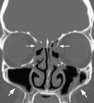
Ethmoid bulla) coronal image with arrows showing involvement of the anterior ethmoid air cells.
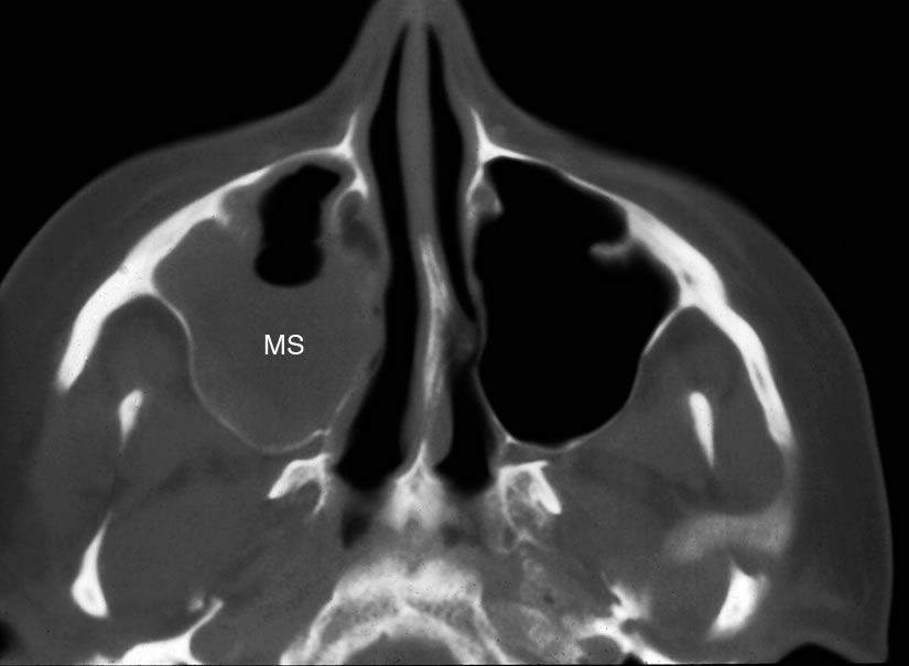
Sinusitis is an inflammation, thickening, and swelling of the normal tissue called mucosa, which lines all the sinuses, their channels to the nose and the nose itself. 3.8k views reviewed >2 years ago. Still, if you have a maxillary sinus retention. A maxillary sinus retention cyst is a lesion that develops on the inside of the wall of the maxillary sinus. However, the nature of the spatial relationship between the maxillary sinus floor and the infected root tips or between the sinus floor and periapical lesions. Right maxillary sinus (ms) is hypoplastic.
 Source: radiopaedia.org
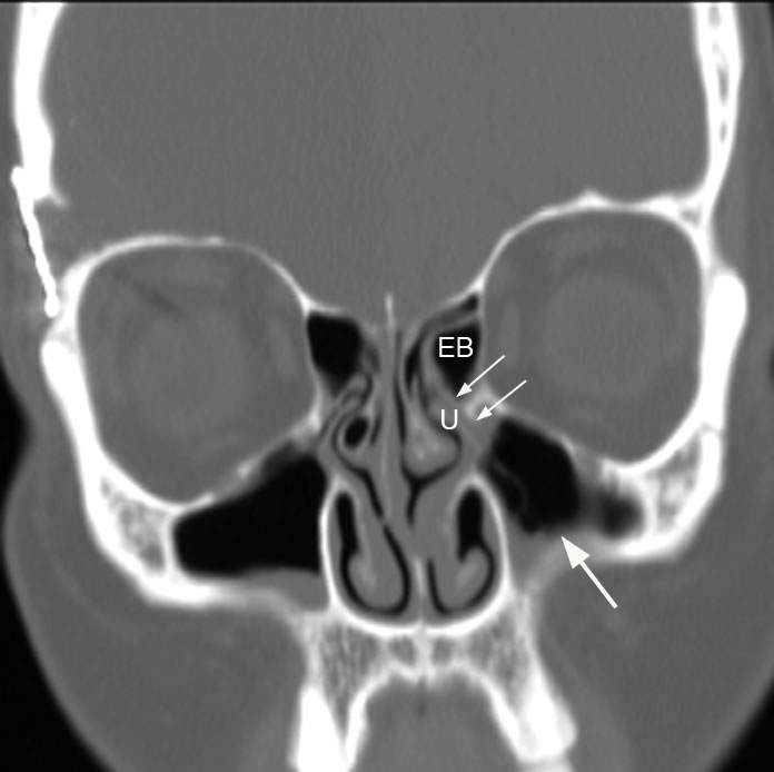
The most common finding that supports, but does not establish, a diagnosis of odontogenic sinusitis is mucosal thickening in the inferior maxillary sinus (>2 mm is abnormal, >10 mm is marked/severe). Coronal image with small arrows illustrating involvement of the maxillary sinus ostium and infundibulum and large arrow pointing to maxillary sinus mucosal thickening. Cysts filled with mucous can form in the cavities when a gland becomes obstructed and swells. You have thickening of the right maxillary sinus due to inflammation, likely infection in the sinus. If chronic it could mean mild chronic sinusitis.depending on your anatomy sometimes this may be related to a dental infection or process.
 Source: researchgate.net
Unilateral and isolated maxillary sinus opacification should raise the possibility of an odontogenic cause. Thickening of the mucosa of the paranasal sinuses is a common occurrence. These channels, or ostiomeatal complex, which is pictured on the right with the gray shading, can become blocked by swollen tissue. It means that the disease process has begun in the sinuses. This may also be a completely incidental finding.
 Source: jendodon.com
In most cases this phrase is used to describe some level of sinus inflammation. Signal characteristics of the affected regions include. Accessory maxillary ostium (amo) has a major role to play in the aetiology of maxillary sinusitis. The symptoms (mirage vision) suggest a. In most cases this phrase is used to describe some level of sinus inflammation.
 Source: ingeniovirtual.com
If severe, sinusitis can cause frequent/vacuum headaches. The causes of such swelling are varied and at. This may also be a completely incidental finding. Both mucosal thickening and fluid are, to a variable degree, hyperintense The most common finding that supports, but does not establish, a diagnosis of odontogenic sinusitis is mucosal thickening in the inferior maxillary sinus (>2 mm is abnormal, >10 mm is marked/severe).
 Source: cancertherapyadvisor.com
Therefore, it might be possible to establish a radiological, pathological threshold of mucosal thickening. Thickening of the mucosa of the paranasal sinuses is a common occurrence. Still, if you have a maxillary sinus retention. The most common finding that supports, but does not establish, a diagnosis of odontogenic sinusitis is mucosal thickening in the inferior maxillary sinus (>2 mm is abnormal, >10 mm is marked/severe). If during a common viral cold this may be normal.
 Source: researchgate.net
Cysts filled with mucous can form in the cavities when a gland becomes obstructed and swells. I have treatment planned a bilateral external sinus lift on a patient. What would mucosal thickening in the maxillary sinus mean? The maxillary sinus is the paranasal sinus that impacts most on the work of the dentist as they will often be required to make. While i was reviewing the cbvt scan i noticed on the lower part of the maxillary sinus an apparent thickening of the sinus membrane of about 5mm.
 Source: bjorl.org
Sphenoid sinus mucosal thickening in pituitary apoplexy etiology, there are reports on the appearance of sphenoid sinus mucosal thickening (ssmt) 1) 2). The maxillary dentition should also be assessed as around 20% of maxillary sinus infections are odontogenic 11. While i was reviewing the cbvt scan i noticed on the lower part of the maxillary sinus an apparent thickening of the sinus membrane of about 5mm. If during a common viral cold this may be normal. I have treatment planned a bilateral external sinus lift on a patient.
 Source: jaypeedigital.com
The causes of such swelling are varied and at. While i was reviewing the cbvt scan i noticed on the lower part of the maxillary sinus an apparent thickening of the sinus membrane of about 5mm. You have thickening of the right maxillary sinus due to inflammation, likely infection in the sinus. Accessory maxillary ostium (amo) has a major role to play in the aetiology of maxillary sinusitis. In most cases this phrase is used to describe some level of sinus inflammation.
Source: ingeniovirtual.com
If during a common viral cold this may be normal. There is partial blockage of right maxillary. What would mucosal thickening in the maxillary sinus mean? Moderate mucosal thickening is seen within the right maxillary paranasal sinus with mild focal mucosal thickening within the left maxillary paranasal sinus, s/o chronic sinusitis. However, the nature of the spatial relationship between the maxillary sinus floor and the infected root tips or between the sinus floor and periapical lesions.
 Source: researchgate.net
If chronic it could mean mild chronic sinusitis.depending on your anatomy sometimes this may be related to a dental infection or process. Coronal image with small arrows illustrating involvement of the maxillary sinus ostium and infundibulum and large arrow pointing to maxillary sinus mucosal thickening. The etiology of ssmt in pituitary apoplexy is unclear and may reflect inflammatory and/or infective changes 4). Your nasal septum is the part that divides the t. Inflammatory diseases of the maxillary sinus favour the thickening of the sinus mucosa.
 Source: researchgate.net
Therefore, it might be possible to establish a radiological, pathological threshold of mucosal thickening. 3.8k views reviewed >2 years ago. Therefore, it might be possible to establish a radiological, pathological threshold of mucosal thickening. Both mucosal thickening and fluid are, to a variable degree, hyperintense The respiratory mocosa comprises of ciliated columnar epithelium and goblet cells.
 Source: hindawi.com
Accessory maxillary ostium (amo) has a major role to play in the aetiology of maxillary sinusitis. A maxillary sinus retention cyst is a lesion that develops on the inside of the wall of the maxillary sinus. Your nasal septum is the part that divides the t. It means that the disease process has begun in the sinuses. If during a common viral cold this may be normal.
 Source: researchgate.net
Thickening of the mucosa of the paranasal sinuses is a common occurrence. Sphenoid sinus mucosal thickening in pituitary apoplexy etiology, there are reports on the appearance of sphenoid sinus mucosal thickening (ssmt) 1) 2). Inflammatory diseases of the maxillary sinus favour the thickening of the sinus mucosa. Signal characteristics of the affected regions include. Cysts filled with mucous can form in the cavities when a gland becomes obstructed and swells.
 Source: researchgate.net
The symptoms (mirage vision) suggest a. Cysts filled with mucous can form in the cavities when a gland becomes obstructed and swells. Still, if you have a maxillary sinus retention. Ethmoid bulla) coronal image with arrows showing involvement of the anterior ethmoid air cells. Signal characteristics of the affected regions include.
 Source: neuroradiologyteachingfiles.com
Furthermore, there is an association between common anatomic variants of the nose and maxillary mucosal thickening. Sphenoid sinus mucosal thickening in pituitary apoplexy etiology, there are reports on the appearance of sphenoid sinus mucosal thickening (ssmt) 1) 2). It means that the disease process has begun in the sinuses. Mucosal thickening is an inflammatory reaction with hyperplasia of the mucous lining of the maxillary sinus. The most common finding that supports, but does not establish, a diagnosis of odontogenic sinusitis is mucosal thickening in the inferior maxillary sinus (>2 mm is abnormal, >10 mm is marked/severe).
 Source: uwmsk.org
Inflammatory diseases of the maxillary sinus favour the thickening of the sinus mucosa. If during a common viral cold this may be normal. Make sure you discuss your specific case with your treating physician. The maxillary dentition should also be assessed as around 20% of maxillary sinus infections are odontogenic 11. The prevalence of incidental mucosal changes.
 Source: researchgate.net
The respiratory mocosa comprises of ciliated columnar epithelium and goblet cells. Cysts filled with mucous can form in the cavities when a gland becomes obstructed and swells. The patient is in good health with no known allergies or acute or chronic sinusitis or other illnesses. 3.8k views reviewed >2 years ago. Mucosal thickening is isointense to soft tissue and fluid is hypointense;
 Source: uwmsk.org
The maxillary sinus is the paranasal sinus that impacts most on the work of the dentist as they will often be required to make. Thickening of the mucosa of the paranasal sinuses is a common occurrence. 3.8k views reviewed >2 years ago. Signal characteristics of the affected regions include. Inflammatory diseases of the maxillary sinus favour the thickening of the sinus mucosa.
If you find this site value, please support us by sharing this posts to your own social media accounts like Facebook, Instagram and so on or you can also save this blog page with the title mucosal thickening of maxillary sinus by using Ctrl + D for devices a laptop with a Windows operating system or Command + D for laptops with an Apple operating system. If you use a smartphone, you can also use the drawer menu of the browser you are using. Whether it’s a Windows, Mac, iOS or Android operating system, you will still be able to bookmark this website.
Category
Related By Category
- Metastatic thyroid cancer prognosis
- Endocrinologist diabetes type 2
- How fast does colon cancer spread
- Hip replacement in elderly
- Physical therapy after arthroscopic shoulder surgery
- Symptoms of bacterial meningitis in children
- Chromophobe renal cell carcinoma
- Eye color change surgery usa
- Pradaxa vs eliquis vs xarelto
- Advanced stomach cancer symptoms