Thyroid cancer ultrasound colors
Home » Doctor Visit » Thyroid cancer ultrasound colorsThyroid cancer ultrasound colors
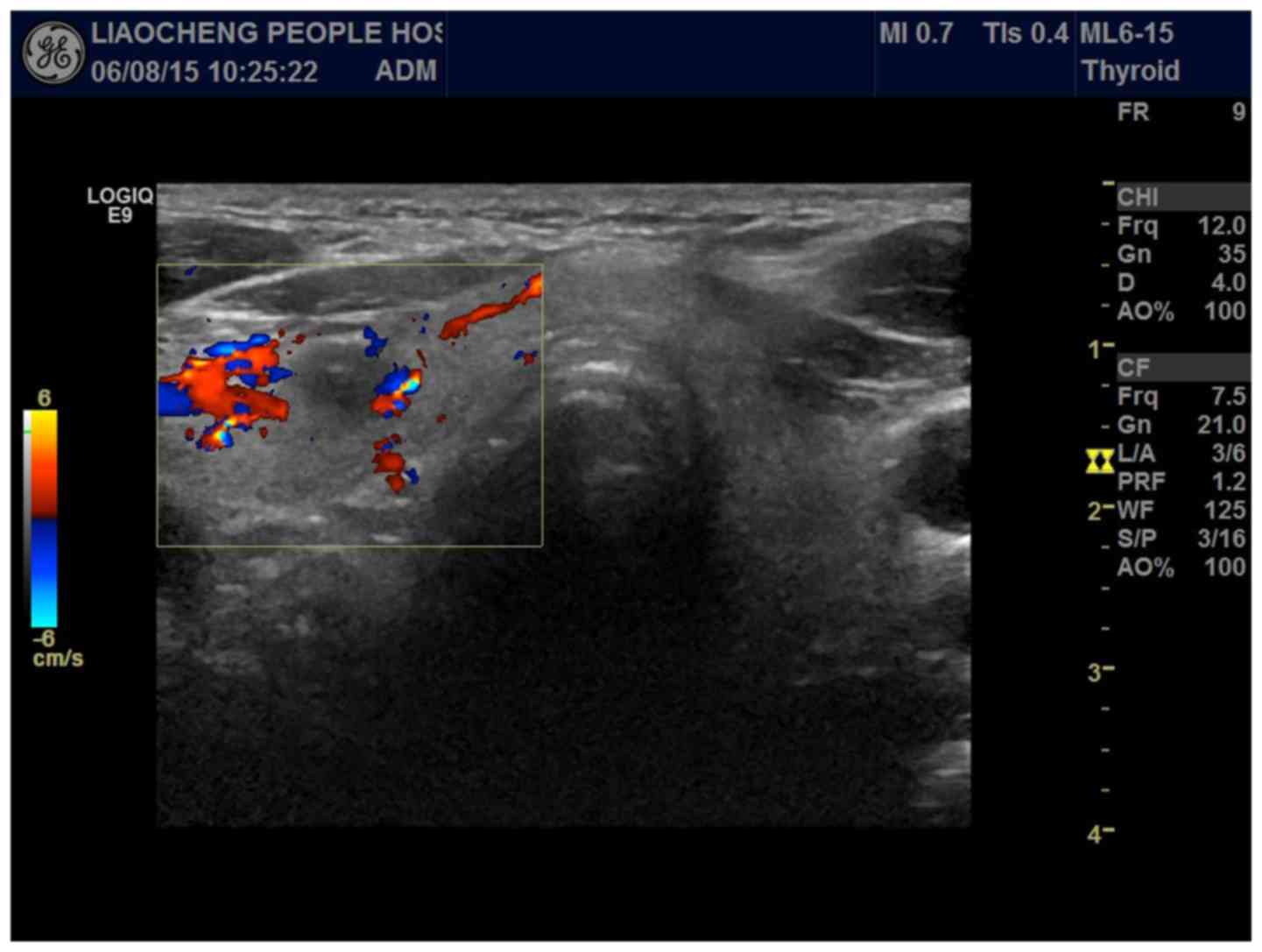
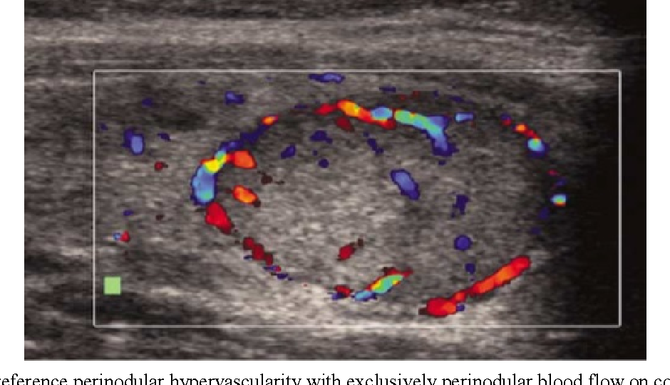
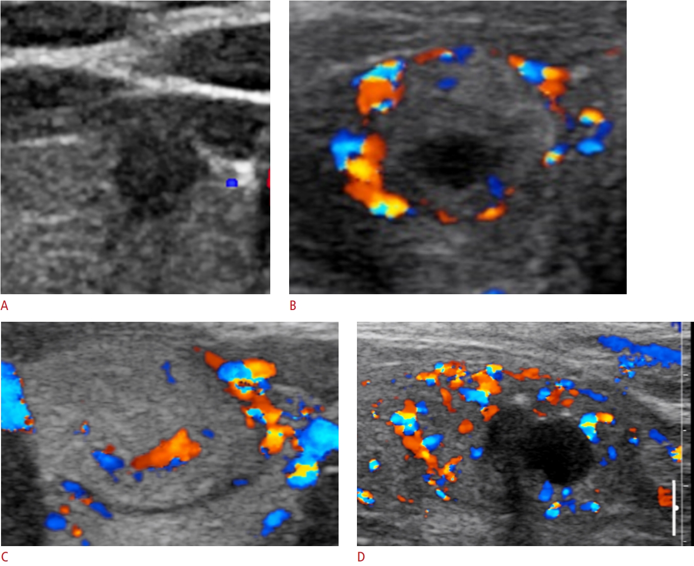
Thyroid Cancer Ultrasound Colors. Ultrasound images thyroid nodule color doppler echogramm 463 pin on thyroide a gallery of high resolution ultrasound color doppler 3d images thyroid related. Color on your thyroid ultrasound means that color doppler was applied and blood flow was detected. The staging system was developed by the american joint committee on cancer (ajcc) and is called the tnm system. Several reports have proposed that increased vascular flow on color doppler sonography may be associated with malignancy in thyroid nodules.
 Role Of Color Doppler Us. (A) Transverse Gray-Scale Image Of… | Download Scientific Diagram From researchgate.net
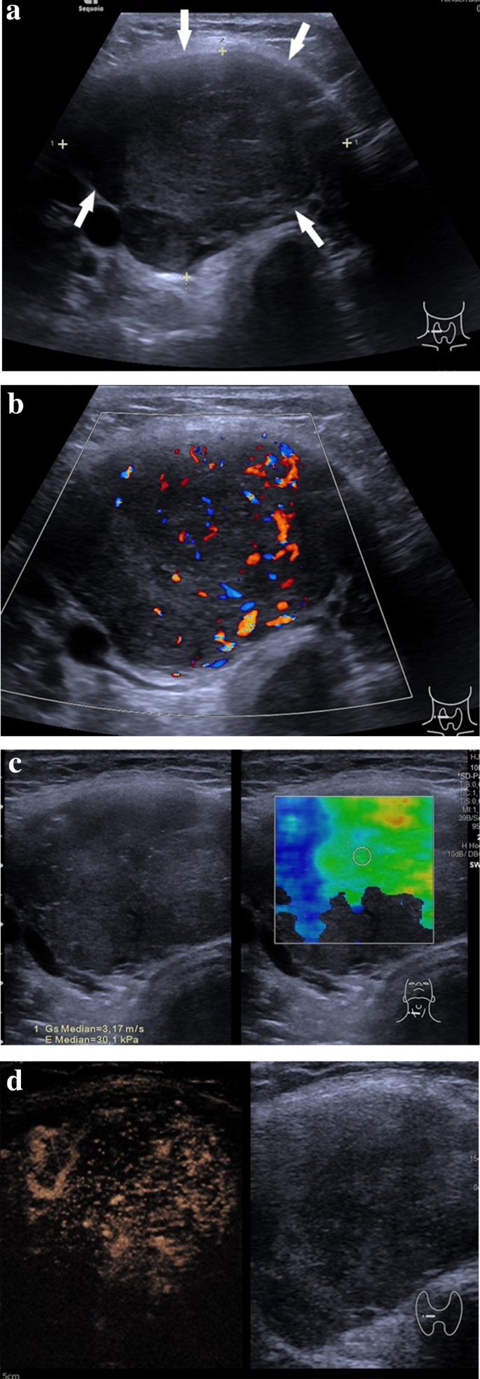
Another common test is the ultrasound. Thyroid disorder grayscale ultrasound color doppler key features; Ultrasound images thyroid nodule color doppler echogramm 463 pin on thyroide a gallery of high resolution ultrasound color doppler 3d images thyroid related. Color on your thyroid ultrasound means that color doppler was applied and blood flow was detected. The color green indicates the median stiffness. Jun 29, 2019 · breast changes include benign conditions and those that increase the risk of breast cancer.
By definition, the flow towards the transducer is represented in red while the flow away from the transducer is represented in blue.
A thyroid ultrasound is a safe, painless procedure that uses sound waves to examine the thyroid gland. This simple test uses sound waves to image the thyroid. Others have described no correlation between the presence of flow and risk of malignancy. Ultrasonography can deliver a diagnostic accuracy of over 90% in thyroid carcinoma, especially papillary carcinoma. Thyroid nodules were found in 97% of patients with thyroid cancer and in 56% of without thyroid cancer. Monitor thyroid cancer patients for residual disease or early evidence of reappearance of malignancy in the thyroid bed or lymphadenopathy, 11.
 Source: onlinelibrary.wiley.com
On average, 1 case of thyroid cancer was found for every 111 ultrasound exams performed. It accounts for the majority (~70%) of all thyroid neoplasms and 85% of all thyroid cancers 2,4. 5.2k views reviewed >2 years ago. The yellow arrow points to a nodule in the right side of the thyroid gland (the ultrasound pictures are a mirror image: Hypervascularity is a typical finding in people with underlying autoimmune thyroiditis (hashimoto�s or graves disease).
 Source: pinterest.com
• color doppler us uses a computer to convert the doppler measurements into an array of colors. This is often the only test needed. A thyroid ultrasound is a safe, painless procedure that uses sound waves to examine the thyroid gland. On average, 1 case of thyroid cancer was found for every 111 ultrasound exams performed. The 3 mm nodule is likely of.
 Source: e-ultrasonography.org
The yellow arrow points to a nodule in the right side of the thyroid gland (the ultrasound pictures are a mirror image: Staging the tumor helps your doctor determine the best treatment for your thyroid cancer. Monitor thyroid cancer patients for residual disease or early evidence of reappearance of malignancy in the thyroid bed or lymphadenopathy, 11. Several recent studies have demonstrated that both strain elastography and swe predict thyroid cancer risk independently of other ultrasound characteristics. The main symptom of thyroid cancer is a lump or swelling in the front of the neck just.
 Source: spandidos-publications.com
Thyroid ultrasound reveals slight increase in size of lobes from 3 5×1.2x.9 mm to 4.1×2.1x 1.2mm and isthmus is.1. The 3 mm nodule is likely of. Most often it is not detected until it gets to a certain size that would make it physically prominent. It accounts for the majority (~70%) of all thyroid neoplasms and 85% of all thyroid cancers 2,4. A lubricant jelly is placed on the skin.
 Source: researchgate.net
Cystic appearance, hyperechoic punctations / calcifications 2015 american thyroid association. After thyroid cancer is diagnosed, it is staged. This simple test uses sound waves to image the thyroid. Color on your thyroid ultrasound means that color doppler was applied and blood flow was detected. On average, 1 case of thyroid cancer was found for every 111 ultrasound exams performed.
 Source: amazon.com
5.2k views reviewed >2 years ago. Color on your thyroid ultrasound means that color doppler was applied and blood flow was detected. It can be used to help diagnose a wide range of medical conditions affecting the thyroid gland, including benign thyroid nodules and possible thyroid cancers. Another common test is the ultrasound. Thyroid inferno should not be confused with a.
 Source: researchgate.net
Cystic appearance, hyperechoic punctations / calcifications 2015 american thyroid association. The average speed is then converted to a specific color. The yellow arrow points to a nodule in the right side of the thyroid gland (the ultrasound pictures are a mirror image: The image of both the thyroid nodule and the surrounding thyroid tissue can present as red color affecting a large part of the thyroid gland beyond the nodule under investigation. The staging system was developed by the american joint committee on cancer (ajcc) and is called the tnm system.
![Pdf] Ultrasonographic And Color Doppler Ultrasonographic Parameters To Discriminate Thyroid Nodules | Semantic Scholar](https://d3i71xaburhd42.cloudfront.net/ded1eb04d701c6599544f7d895059d61f3a37995/3-Figure1-1.png “Pdf] Ultrasonographic And Color Doppler Ultrasonographic Parameters To Discriminate Thyroid Nodules | Semantic Scholar”) Source: semanticscholar.org
Color on your thyroid ultrasound means that color doppler was applied and blood flow was detected. Thyroid inferno should not be confused with a. Monitor thyroid cancer patients for residual disease or early evidence of reappearance of malignancy in the thyroid bed or lymphadenopathy, 11. Thyroid nodule thyroid cancer ultrasound colors wednesday, august 3, 2022 edit. Ultrasound images thyroid nodule color doppler echogramm 463 pin on thyroide a gallery of high resolution ultrasound color doppler 3d images thyroid related.
 Source: researchgate.net
5.2k views reviewed >2 years ago. Thyroid inferno should not be confused with a. Thyroid nodule thyroid cancer ultrasound colors wednesday, august 3, 2022 edit. Thyroid nodules were found in 97% of patients with thyroid cancer and in 56% of without thyroid cancer. This simple test uses sound waves to image the thyroid.
 Source: youtube.com
It can be used to help diagnose a wide range of medical conditions affecting the thyroid gland, including benign thyroid nodules and possible thyroid cancers. The role of sonography in thyroid cancer. An ultrasound scan of your neck can check for a lump in your if a biopsy finds that you have thyroid cancer, further tests may be needed to check whether the cancer had spread to another part of your body. Thyroid nodules were found in 97% of patients with thyroid cancer and in 56% of without thyroid cancer. This is often the only test needed.
 Source: sites.google.com
Increased color doppler flow is suspicious. Cystic appearance, hyperechoic punctations / calcifications 2015 american thyroid association. A thyroid ultrasound is a safe, painless procedure that uses sound waves to examine the thyroid gland. It accounts for the majority (~70%) of all thyroid neoplasms and 85% of all thyroid cancers 2,4. The main symptom of thyroid cancer is a lump or swelling in the front of the neck just.
 Source: semanticscholar.org
The fna will usually (but not always) tell if a nodule is benign or malignant. Another common test is the ultrasound. Thyroid nodules were found in 97% of patients with thyroid cancer and in 56% of without thyroid cancer. However, a cystic lymph node in the bottom half of the neck is most commonly a diagnosis of papillary thyroid cancer. Thyroid nodules were found in 97% of patients with thyroid cancer and in 56% of without thyroid cancer.
 Source: e-ultrasonography.org
Hypervascularity is a typical finding in people with underlying autoimmune thyroiditis (hashimoto�s or graves disease). Thyroid disorder grayscale ultrasound color doppler key features; The risk of cancer increased with the size of. The image of both the thyroid nodule and the surrounding thyroid tissue can present as red color affecting a large part of the thyroid gland beyond the nodule under investigation. However, a cystic lymph node in the bottom half of the neck is most commonly a diagnosis of papillary thyroid cancer.
 Source: link.springer.com
It accounts for the majority (~70%) of all thyroid neoplasms and 85% of all thyroid cancers 2,4. This simple test uses sound waves to image the thyroid. A thyroid lobe has the shape of the rotation ellipsoid. Thyroid ultrasound reveals slight increase in size of lobes from 3 5×1.2x.9 mm to 4.1×2.1x 1.2mm and isthmus is.1. Thyroid nodules were found in 97% of patients with thyroid cancer and in 56% of without thyroid cancer.
 Source: clinicalimaging.org
The 3 mm nodule is likely of. The green arrow points to the breathing tube (trachea). However, a cystic lymph node in the bottom half of the neck is most commonly a diagnosis of papillary thyroid cancer. It accounts for the majority (~70%) of all thyroid neoplasms and 85% of all thyroid cancers 2,4. In our experience, most benign thyroid nodules are either purple or green.
 Source: researchgate.net
Increased color doppler flow is suspicious. Thyroid nodule thyroid cancer ultrasound colors wednesday, august 3, 2022 edit. It accounts for the majority (~70%) of all thyroid neoplasms and 85% of all thyroid cancers 2,4. However, a cystic lymph node in the bottom half of the neck is most commonly a diagnosis of papillary thyroid cancer. The staging system was developed by the american joint committee on cancer (ajcc) and is called the tnm system.
 Source: youtube.com
It can be used to help diagnose a wide range of medical conditions affecting the thyroid gland, including benign thyroid nodules and possible thyroid cancers. The average speed is then converted to a specific color. In our experience, most benign thyroid nodules are either purple or green. The risk of cancer increased with the size of. Thyroid nodules were found in 97% of patients with thyroid cancer and in 56% of without thyroid cancer.
 Source: umbjournal.org
A thyroid lobe has the shape of the rotation ellipsoid. The risk of cancer increased with the size of. The purpose of this study was to determine whether the vascularity of a thyroid nodule can aid in the. The average speed is then converted to a specific color. Color on your thyroid ultrasound means that color doppler was applied and blood flow was detected.
If you find this site value, please support us by sharing this posts to your own social media accounts like Facebook, Instagram and so on or you can also save this blog page with the title thyroid cancer ultrasound colors by using Ctrl + D for devices a laptop with a Windows operating system or Command + D for laptops with an Apple operating system. If you use a smartphone, you can also use the drawer menu of the browser you are using. Whether it’s a Windows, Mac, iOS or Android operating system, you will still be able to bookmark this website.
Category
Related By Category
- Metastatic thyroid cancer prognosis
- Endocrinologist diabetes type 2
- How fast does colon cancer spread
- Hip replacement in elderly
- Physical therapy after arthroscopic shoulder surgery
- Symptoms of bacterial meningitis in children
- Chromophobe renal cell carcinoma
- Eye color change surgery usa
- Pradaxa vs eliquis vs xarelto
- Advanced stomach cancer symptoms